Here's Looking at You, Kid
Click on an image for a larger view
|
I'm quite used to looking at the world using these retinas, and very
unused to seeing what they look like. When I had an episode of an
occluded vein, after a couple of referrals, I ended up at the Doris
Stein eye clinic at UCLA. Lots of tests were done, including having
photographs done of my retinas. When I asked the photographer if I
could have copies of the photos, she asked if I wanted TIFF or JPEG.
TIFFs, of course! I've done some enhancement to make details stand
out better, and what you'll see when you click is a high quality JPEG.
|
 |
 |
|
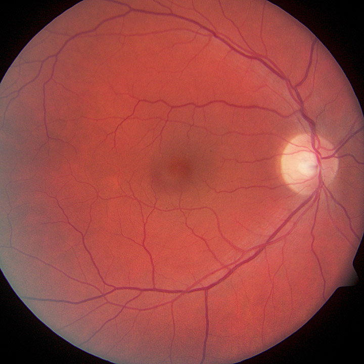
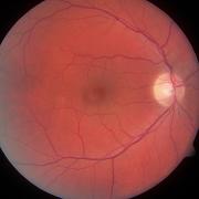
My left eye's retina, in living color
|
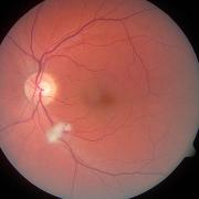
This one must be the right eye
|
|
A bit of explanation is in order. The bright spots on the left and
right in the image pairs are the optic nerves. These are the conduits
through which images are seen, and blood enters & leaves the eye. The
center of vision is in the center of most of the photos. Veins are
darker and generally larger than arteries. I like the way in the
right eye where the vein and artery twist around each other. Who did
that?
|
 |
 |
|
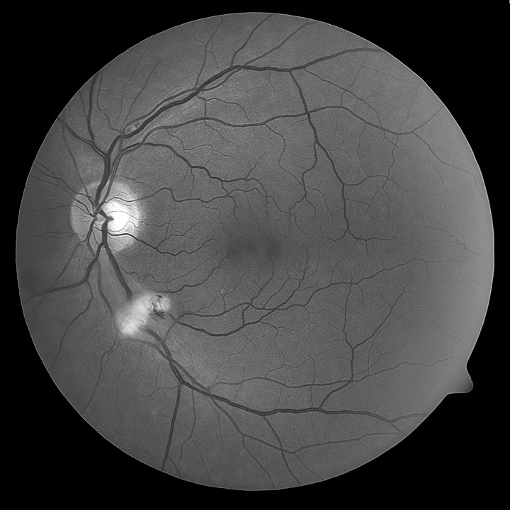
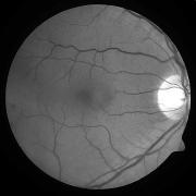
Left eye in B&W. The bleeding is at 8 o'clock
|
Here's that right eye - normal. Note there are no blood vessels in the center of vision
|
 |
 |
|
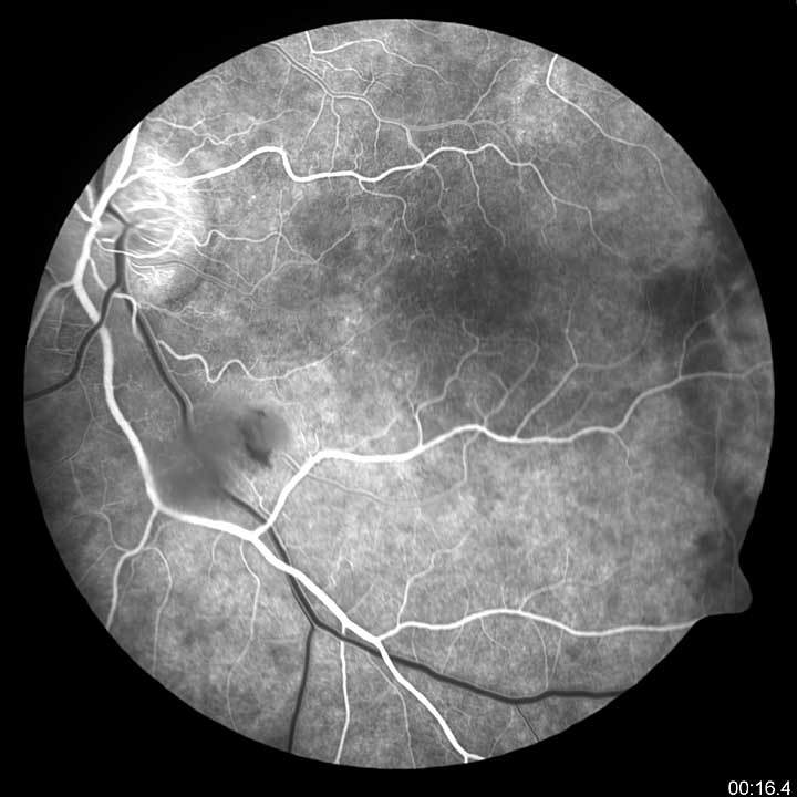
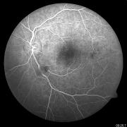
The fluorescein dye enters the left eye. Now the (smaller) arteries are white and veins are black
|
The dye is now leaving, and the veins are lit up
|